Experiencing leg pain or cramps? It could be Peripheral Artery Disease (PAD) or Knee Arthritis. Learn More

In order to create images of the veins in the body, venous ultrasound uses sound waves. It is widely used, especially in the veins of the leg, to check for blood clots. Searching for blood clots, especially in the leg’s veins, is the most common reason for a venous ultrasound test. They are also known as thrombosis of the deep vein, or DVT.
During ultrasounds, a tiny probe called a transducer and gel are mounted directly on the skin. High-frequency sound waves pass through the body, and photographs are taken in real-time. They can reveal the internal organs of the body's structure and movement as well as the blood flowing into vessels of the blood.
Venous ultrasound gives images of the veins all through the human body. A venous ultrasound examination can include a Doppler ultrasound study.
Some common uses:
In children, venous ultrasound is used to:
Doppler ultrasound images can help doctors evaluate:
Are you looking for an affordable venous ultrasound? At North Atlanta Vascular and Vein Center (Suwanee/Johns Creek, Alpharetta, Cumming and Lawrenceville), we provide safe, accurate, and affordable ultrasounds. For more details, contact- 7707715260
Guidelines to prepare for the ultrasound include:
During the ultrasound, a transducer sends sound waves and records the echoing waves during the exam. It sends small pulses of inaudible, high-frequency sound waves throughout the body as the transducer is pressed against the skin.
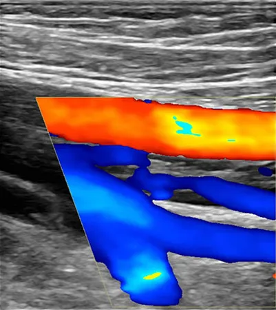
The direction and speed of blood cells as they move through vessels is measured by Doppler ultrasound, a special ultrasound technique. The sounds are collected and processed by a computer and graphs, or colored pictures are produced that represent the blood flow through the blood vessels. This entire procedure is painless.
The procedure is performed in the following steps:
After this procedure, you can expect the following:
The radiologist will analyze the image and send a signed report to the doctor who requested the exam. Your doctor will then communicate the results of the ultrasound with you. A follow-up exam can also be conducted to see if an abnormality has changed over time.